
5
Introduction to the Pelvic Floor
Women are significantly more affected by pelvic health conditions compared to men. Pregnancy and childbirth can lead to a variety of negative outcomes, including urinary incontinence, bowel incontinence, back pain, pelvic organ prolapse, perineal pain, and sexual dysfunction.
To effectively educate patients about the pelvic floor, it is essential to have a clear understanding of its anatomy and physiology. The pelvic floor consists of three layers of fourteen muscles that support our internal organs.
Along with muscles, the pelvic floor consists of crucial ligaments and fascia that support the bladder, reproductive organs, and rectum.
Bones of the Pelvis
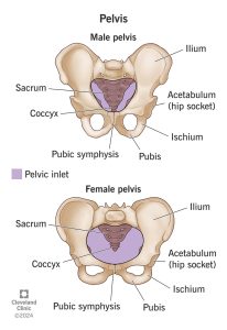 The pelvis consists of the hip bones (ilium, ischium, pubis), along with the sacrum and coccyx.
The male pelvis is narrower and the female pelvis is wider.
Ilium: large, wide bone that takes up the top portion of each half of the pelvis
Ischium: curved bone on the base of each bottom half of the pelvis
Pubis: lowest anterior portion of each half of the pelvis
Sacrum: larger triangular bone located at the base of the spine formed through the fusion of sacral vertebrae
Coccyx: smaller triangular bone below sacrum from fusion of spine
*For the purpose of this Pressbook, I will focus on female anatomy.*
Musculature of the Pelvic Floor
The pelvic floor has three layers of muscles. This chapter will be a brief overview of the three layers and which muscles are in each layer. Next chapter will dive into the functions of the muscles.
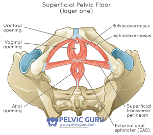
Layer 1: Urogenital Triangle
Bulbocavernosus
Ischiocavernosus
Superficial Transverse Perineal
External Anal Sphincter
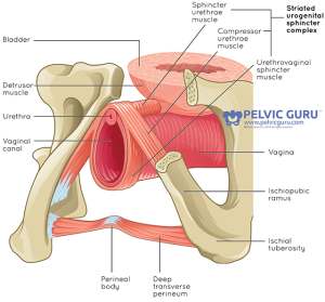
Layer 2: Urogenital Diaphragm
Urethral Sphincter
Compressor Urethrae
Sphincter Urethral Vaginalis
Deep Transverse Perineal
Perineal Membrane
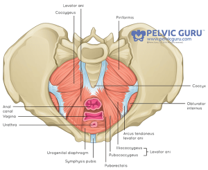
Layer 3: Pelvic Diaphragm
Levator Ani Muscle
-
Pubococcygeus, Pubovaginalis, Puboanalis, Puborectalis, Iliococcygeus
Coccygeus
Piriformis
Obturator Internus
Key Takeaways
- The pelvic floor has three layers of fourteen muscles that support our internal organs.
Explore these courses!
References:
Eickmeyer, S. (2017). Anatomy and physiology of the pelvic floor. Physical Medicine and Rehabilitation Clinics of North America, 28(3), 455-460. https://doi.org/10.1016/j.pmr.2017.03.003
Jones, J., Domanico, J., Peek, H., Lee, T. E., & Kern, L. A. (2020). Promoting women’s health and wellness. American Occupational Therapy Association. https://www.aota.org/publications/ot-practice/ot-practice-issues/2020/womens-health
Physiopedia. (2020, October 17). Pelvic floor anatomy. https://www.physio-pedia.com/Pelvic_Floor_Anatomy
Picture reference:
Cleveland Clinic. (2024, September 24). Pelvis: What is it, where is it, types & anatomy. https://my.clevelandclinic.org/health/body/pelvis
Pelvic Global Academy. (2024). https://pelvicglobal.com/beginners-guide-to-pelvic-floor-muscle-exercises/
Media Attributions
- pelvis
- Art29-copy
- Art30-copy
- Art3-copy